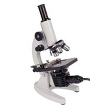
Microscopy
Here is my short guide to buying and using a Microscope! Unlike Telescopes and binoculars, some (though not all!) cheap microscopes sold as toys can have a good optical performance and actually be used at their highest advertised magnification. CCD microscopes that display the image onscreen on the VDU are very good and often very reasonable pricewise, though quite often (but not always!) use lower magnifications. The top end is binocular microscopes but these can be expensive, though an excellent quality instrument can be bought for about £130. For studying small cells such as bacteria, up to 2000x magnification is needed, but for general studies, 80x is adequate. The magnification is the product of the eyepiece x objective, so a 20x eyepiece and 100x objective would give a 2000x magnification. It is very advisable to have a built in light source, preferably mains operated, especially for very high magnifications. The highest magnifications are with electron microscopes but these are highly professional and inordinately expensive instruments!
You don’t have to spend a large amount to get a usable microscope if you know what to look for. Children over 8 years old can get a really good introduction to the hobby for a relatively cheap birthday or Christmas present. There are some duds out there in the department stores, better to buy from a toyshop or store where they will let you try one out first to see and make sure it has good usable optical quality and a non-flimsy and easy to adjust focusing mechanism! Be sure to try it at high as well as low magnification!
For a starter microscope, for hobby use, the Microscope Lab microscope as supplied eg by Edu Science, Elenco, Discovery Planet, Discovery World and others, in their Microscope Lab Max kit (for just £12- £25), is excellent, and eminently usable even at 900x. The image is excellent at lower magnifications, and very good at 900x (or 1200x depending on the model). The 900x model has 8x, 25x, and 50x objective lenses, with a Huygens' Eyepiece with 12x to 18x zoom, giving magnification ranges of 96x-144x, 300x-450x and 600x-900x. There is a colour filter and diaphragm wheel with a battery bulb light and a mirror. They contain most of what’s needed to start with in the kits. This includes a Petri dish, safety scalpel, slides and covers (though the covers are plastic), collecting vials, and some basic reagents/ colouring agent. You can easily add test tubes and culture dishes for reagents and cultures. The microscope supplied is a sophisticated and capable scientific instrument, especially considering its extremely reasonable price!
Do be aware that cultures eg moulds, bacteria made in limited oxygen conditions can often be harmful (ie poisonous) and a possible serious risk in breathing dust from without a breathing mask.
Basically you can have ‘microscopes’ with a low power eyepiece and objective for viewing larger items at lower magnifications (up to about 80x) where the specimen can be manipulated while viewing, or more powerful ‘Compound’ microscopes with higher power eyepieces and particularly higher power objective lens systems that need prepared slides. It is very good to have one of each sort!!!! Especially as compound microscopes usually start at 100x magnification upwards! A modest 20x -40x illuminated magnifier can be bought for well under £10! There are also stereo (binocular) microscopes with eg 40 -80x magnification that give stunning 3D views of larger specimens, though you might have to be very lucky to get one of these for under £50. Do try car boot sales for these!
Incidentally, regarding magnification, many smaller medical labs only use up to 400x, though some of the bigger facilities, with rarer conditions to investigate may use 1000x - 1500x, so some of the cheaper microscopes that provide 900x - 1200x maximum are actually powerful and capable instruments!
The higher the magnification, the more difficult it is to focus; light adequately and find the part of the slide to view. Very high magnifications for optical microscopes have an extremely small field of view. They also have very small eye relief, making viewing much more difficult. Generally, compound microscopes have a set of different power objectives on a rotatable turret, to let lower powers and hence lower magnifications to be used while finding different parts of specimens by moving the slide. They may also have interchangeable eyepieces for further adjustment of the magnification. Focusing is much trickier and more difficult to achieve with higher powers, and the objective lens is very close indeed to the slide.
It is necessary to re-focus the microscope each time the magnification or specimen slide is changed. As the depth of field in focus is very small, it may be necessary to re-focus when moving the specimen slide. Focusing and lighting are the two most important factors apart from slide preparation. You can't take too much trouble and care in getting these right, and both often need a very fine light touch to get just right!
Some specimens are ‘light shy’ and need a means to adjust the brightness of the illuminating lamp. This can be tricky to achieve with cheaper microscopes. Mirror light illumination system can be adjusted for brightness by moving the mirror. Battery systems will need some kind of filter or ‘iris’ system and variable transformers (or eg dimmer switches) or more complex iris systems are needed with mains lighting systems. Bulbs are easier to adjust, as modern LCD’s and halogen spotlights are very bright, though it may be possible to partly dim halogen spotlights though reducing their life. Microscopes with a condenser lens may need the condenser position carefully adjusted for best lighting effect.
A very useful tip for specimen viewing is that even different parts of the same slide nearly always benefit from adjusting the light level each time the slide is moved. Always re-centre the part of the specimen you wish to view under a higher magnification! Never use direct sunlight on a microscope mirror for illumination, as a very important safety measure. Always set the objective lens approximately in position by side-ways viewing, before looking through the eyepiece and focusing. Mineral water or distilled water is better to make wet mounts for slides as tap water can kill small organisms because of its chlorine (and fluorine, if present) content. Many specimens are transparent and benefit greatly from staining with coloured dyes.
More powerful optical compound microscopes use oil immersion objectives, where a drop of fine special oil is used to fill the usual air gap between the objective lens and the specimen slide. This gives a clearer image at magnifications above about 1200x. More expensive microscopes also have a fine tuning mechanism for higher magnifications, and a mechanical slide stage to move the specimen very small amounts horizontally right to left and up or down.
Cheap Indian inks, in a range of colours when diluted can be used to stain home made specimen slides. To prepare a home slide, a fine scalpel or safety razor blade is used. A lit stand magnifier will help enormously, or use at least +4.00 diopter glasses to see clearly, using a cutting board, and be especially careful not to slice your fingers,. Generally, the finer the thinness of the specimen slice, the better. It may take several (or many!) attempts to get a really good specimen eg from a slice of a leaf or a plant stem. If necessary, the specimen is then stained, and mounted on a slide with a thin glass cover slip attached around its edges lowered one side first to avoid air bubbles, and ‘fixed’ in place around the edges . For liquid specimens, again staining will usually help greatly, and a very small amount is pipetted into a special slide with a small dish curved into it, and again a cover slip is added. It helps very greatly to completely avoid fingerprints on the slide or cover disk, unless you wish to view fingerprints!!!! I once made a really excellent slide from the cross-section of a plant stem in this way, it can just take some practice! A needle and fine tweezers are essential for handling small specimen slices. An eye dropper pipette is very useful for putting one small drop of a liquid or solution onto a slide. You can also drop some melted candle wax onto the specimen and when set, this makes cutting a fine slice much easier. If this is done in the head of a nut protruding partly from a bolt, the nut can be unscrewed very finely and a scalpel used to cut a layer (eg of plant material) maybe just one cell thick - ideal as a specimen for the microscope!
Cover slips or cover glasses are a very thin squareor round piece of glass (or plastic) that is placed over a water drop on the slide. Because of surface tension, a water drop sits as a thick dome. With a cover slip in place, the drop is flattened out by capillary attraction allowing focusing with high power very close to the specimen. The cover glass also confines the specimen to a single plane and thereby reduces the amount of focusing necessary. Finally, the cover glass protects the objective lens from immersion into the water drop and provides permanent protection for a mounted slide. With an oil immersion objective, it allows a drop of specially refractive oil to be held between the objective lens and the cover glass, and keeping oil away from the specimen.
Some thicker samples need pieces of cover glass to build up a thicker channel before the cover glass top layer is added. With fast moving aquatic specimens, either add some cotton or cotton wool strands to confine their movement, or add a very small drop of alcohol or washing up liquid or very dilute bleach to slow them down. A commercial go slow preparation can be bought, which slows them down for ten minutes or so very well.
To prepare specimens from living matter, that will last fairly permanently, the routine is to put them into hot (not boiling) 70% alcohol, transfer when cool into 90% -95% alcohol for some hours, set in clove oil and then put onto a slide coated with some balsam. If this preparation is too much (you are unlikely to convince your pharmacist to sell you neat 100% proof alcohol), then for semi-permanent preservation, put into some hot vodka (DON’T overheat, it will flame! And never leave unattended!), leave to cool and stand eg overnight or for 24 hours, allow the alcohol to evaporate and fix on the slide with either some hairspray or gum Arabic. Don’t drink any leftover Vodka if it has been tainted with biological specimens! Alternatively, put some 40% Vodka in the freezer (not 37% proof, this freezes!) and freeze dry the specimen and mount as before with hairspray or gum Arabic, before adding a cover slip. This won’t work for some specimens where ice crystal damage can occur while freezing. In any case, the Vodka is improved for drinking by this treatment! (leaving it in the freezer, not soaking specimens!) Children need parental supervision in preparing slides. If fingers are cut while handling eg soil samples or some biological specimens, medical advice should be urgently sought.
Sets of pre-prepared slides are available and are usually of a very high quality. Some can be imported from the USA at eg 100 slides for £40 including postage. Some UK prepared sets are very expensive by comparison. Secondhand shops or car boot sales can often provide real bargains here, though the quality of the slides can be variable. Catalogues of the larger sets can provide good ideas for specimens to prepare for yourself!
Small compound hand magnifiers are useful to use ‘in the field’, and some have a chamber to hold specimens for easy viewing and to hold live specimens temporarily!
Hand magnifiers eg jeweller’s loupe type often have the magnification quoted as an area magnification where the 30x actually means just over 5x size magnification.
Small insects and insect parts, butterfly parts, pond wildlife (eg algae and parasites in pond water), seeds, pollen, plants and trees, flower petals, mosses and lichens, minerals and crystals, moulds and fungi, bird feathers, small cellular organisms, fish parts, natural and artificial fibres, biological specimens(eg skin, hair, tissue, blood) and suchlike all make very good slide specimens if prepared properly.
Be very careful when focusing at higher powers, not to hit/ break the slide cover (or slide!), as it also very likely in so doing to damage or break the objective lens.
You can be amazed at what you can see even in a quality budget microscope. As an example of the extent of the microscopic world, there are more living, tiny organisms in a tablespoon of soil (though you may only be lucky enough to see some of the larger ones!) than there are people on Earth! The detail in many microscope specimens is truly remarkable. If you have a scanner, try scanning a portion of a pencil line at a high resolution and viewing the resulting image at a higher magnification. This scanner technique will also work for a range of specimens, eg cloth etc.
One very useful accessory is a polarising filter, which helps cut down reflections depending on the angle it is turned around to. Double polarising filters are good at setting the amount of light used to illuminate the specimen. By varying the rotation of the two filters, fine adjustment to the illumination is possible. By putting one filter behind the specimen and one in front (at a lower magnification!) stress patterns can be observed eg in clear plastics!
Maintenance consists of cleaning lenses outside surfaces, with a clean soft cloth and neat alcohol, or lens cleaning fluid. If necessary, a tiny drop of oil or very small amount of white or copper grease can be very carefully put on the focusing mechanism. That’s it, nothing else! And never try to dismantle the microscope, ever!
Protect the microscope from dust when not in use and it will give many, many years of faithful service!! If kept in a warmer dry location, problems with mould and fungal growth will also be avoided.
Please don’t drop or force mechanisms! Modern microscopes are precision instruments that have been developed to a high degree of mechanical and optical perfection over very many years. While the microscope presents an image, a great part of the skill is in observing and interpreting what is seen. A lot of this comes down to practical experience, theoretical knowledge and sometimes a great deal of actual practice, and particularly, a great deal of patience and very careful observation!
Do read a great deal, eg identifying the main types of parasites in pond or aquarium water is a lot easier if you have some idea what you are looking for! There are many good sites on the Internet! Look for the specimen type and the word microscope in eg Google!
Microscopy can be a very entertaining and enjoyable hobby ! (and eg for aquarium keepers an essential aid and diagnostic tool).
You can also read articles on the Internet telling you about the techniques that are used by professionals but which are beyond the scope of amateur investigations! You may be able to take digital photographs as well through the microscope, but focusing the camera may partly be trial and error!
This article doesn’t cover more advanced aspects of microscopy ie,Phase-Contrast and Dark Field Microscopy; Fluorescence Microscopy; Polarising and Interference Contrast Microscopy; Microphotography; Electron Microscopy and Preparation Techniques for Electron Microscopy. There suitable articles on the Internet if you wish to read more on any of these topics. It is interesting to find out how they achieve results that are difficult or impossible by other means, even if the practical realm for most of these is the world of professional microscopists.